

WHAT IS PAMO?
A Non-Surgical Medical Procedure
for Selected Adolescents with Idiopathic Scoliosis
A standardized, hospital-based procedure.
PAMO (Paraspinal Muscle Optimization) is a standardized medical procedure developed for selected adolescents with idiopathic scoliosis. Performed under image guidance in a hospital environment, the procedure was developed following years of clinical observation, anatomical investigation, imaging analysis, and protocol development.
PAMO is currently performed exclusively by Professor Semih Keskil in Ankara, Türkiye.
PAMO is performed under image guidance in a hospital environment without incisions, stitches or general anesthesia.
Nearly a decade of development, built on four decades of experience.
PAMO did not emerge from a single research project or laboratory concept. Its origins lie in more than four decades of clinical experience in pediatric neurosurgery and spinal disorders.
Throughout his career, Professor Semih Keskil treated numerous adolescents with scoliosis and observed a recurring challenge faced by patients, families, and physicians alike. While bracing and surgical correction remain important components of scoliosis management, many patients struggled with long-term brace compliance, and many families were reluctant to consider major spinal surgery unless absolutely necessary.
"Is there another option?"
These observations led Professor Keskil to explore a fundamental clinical question:
Could asymmetries within the paraspinal muscles play a greater role in scoliosis progression than previously thought?
Over the following years, this question became the focus of extensive anatomical study, radiological review, clinical observation, and procedural development. The result was the creation of a standardized hospital-based procedure designed to address selected muscular structures associated with spinal support and balance.
Today, PAMO represents the culmination of nearly a decade of focused development built upon more than forty years of clinical experience.
Why paraspinal muscle balance matters.
The development of PAMO was based on the observation that asymmetry within the paraspinal musculature may play an important role in the progression of selected cases of adolescent idiopathic scoliosis.
This observation led to further investigation into the relationship between muscular balance, spinal alignment, and curve progression. Over time, these investigations contributed to the development of a standardized clinical protocol designed to address specific muscular structures associated with spinal support and function. PAMO was developed within this clinical and anatomical framework.
The Role of Paraspinal Muscles
The paraspinal muscles play an important role in spinal alignment through the regulation of muscle tone, force symmetry, and functional control. When these mechanisms become imbalanced, asymmetric forces may be transmitted to individual vertebral segments. Over time, these forces may contribute to the maintenance and progression of spinal curvature in selected patients.
The development of PAMO was based on the clinical observation that muscular asymmetry may represent more than a secondary consequence of scoliosis. In selected patients, imbalances within the paraspinal musculature may contribute to the rotational and lateral forces acting upon the spine.
This perspective led Professor Semih Keskil to investigate whether selected muscular structures could represent a meaningful point of clinical intervention. PAMO was developed within this clinical framework. Performed under image guidance in a hospital environment, the procedure focuses on restoring functional balance within selected paraspinal muscle groups rather than addressing spinal curvature alone.
The objective is to support spinal balance, alignment, and function through a procedure designed specifically for carefully selected patients with adolescent idiopathic scoliosis.
PAMO focuses on restoring functional balance within selected paraspinal muscle groups rather than addressing spinal curvature alone.
Adolescents, 10–20 years, Cobb angle 20°–50°
With ongoing skeletal growth. Final eligibility is determined after comprehensive evaluation in Ankara.
Adult, congenital, or neuromuscular scoliosis
Patients with completed skeletal maturity, structural vertebral abnormalities, or underlying neurological/muscular disorders.
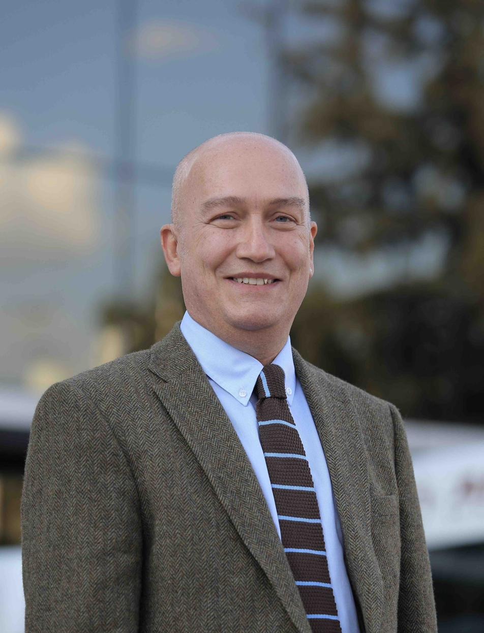
Pediatric neurosurgeon, researcher, developer of PAMO.
Still Have Questions?
Find answers to the questions families ask most frequently about PAMO.
View Frequently Asked QuestionsReady to Learn More About PAMO?
A free remote preliminary assessment is the first step in understanding whether PAMO may be appropriate for your child.